ADHD
Amino acids and ADHD: why the studies give no answer
14 September 2026 · By Dr. B.J. Huber · 42 min read
Dopamine is built from tyrosine, serotonin from tryptophan. From this follows an obvious question: are these building blocks missing in ADHD, and if so, which ones? Online there are very clear answers. Amino acid profiles are offered as diagnostics, tyrosine and taurine are recommended as supplements, and single percentages travel from one page to the next.
I looked at the work behind this, every single study in the original. What emerges has less to do with your values than with the way those values were measured. In this article we look at what was actually studied, why most findings vanish when you recalculate, and why the existing studies probably cannot answer your question at all.
- There is no established amino acid deficit in ADHD. The only study that calculated correctly finds nothing.
- The reason lies in the measurement method. An amino acid profile captures 22 values at once, and one or two abnormal values per study are expected by chance alone.
- The entire profile literature comes from children. A complete plasma amino acid profile in adults with ADHD does not exist.
- Where adults have been measured, the direction reverses. Tryptophan is raised in medication-free children and lowered in adults.
- The one finding that holds across all studies is reduced kynurenic acid.
- On taurine in ADHD there is not a single human study.
- As a supplement, too, none of the three well-known amino acids is proven: for tyrosine only depletion has been measured, taurine demonstrably barely reaches the brain, and the glycine sleep studies all come from the manufacturer.
Important first
This article does not say that people with ADHD have no amino acid deficiencies. A deficiency can certainly exist in an individual case, and targeted supplementation can then genuinely help with symptoms. What is missing are clear studies that prove a deficiency typical of ADHD. And a lack of study evidence is not proof that no deficiency exists. If you suspect one, have it measured. Only then the result answers the question about your own value, not the question about ADHD.
Are amino acids missing in ADHD?
A consistent amino acid deficiency typical of ADHD is not established. The largest and methodologically cleanest study found normal values in the blood, in the urine and in protein intake of 83 children with ADHD. The studies that report abnormalities are smaller and calculated differently.
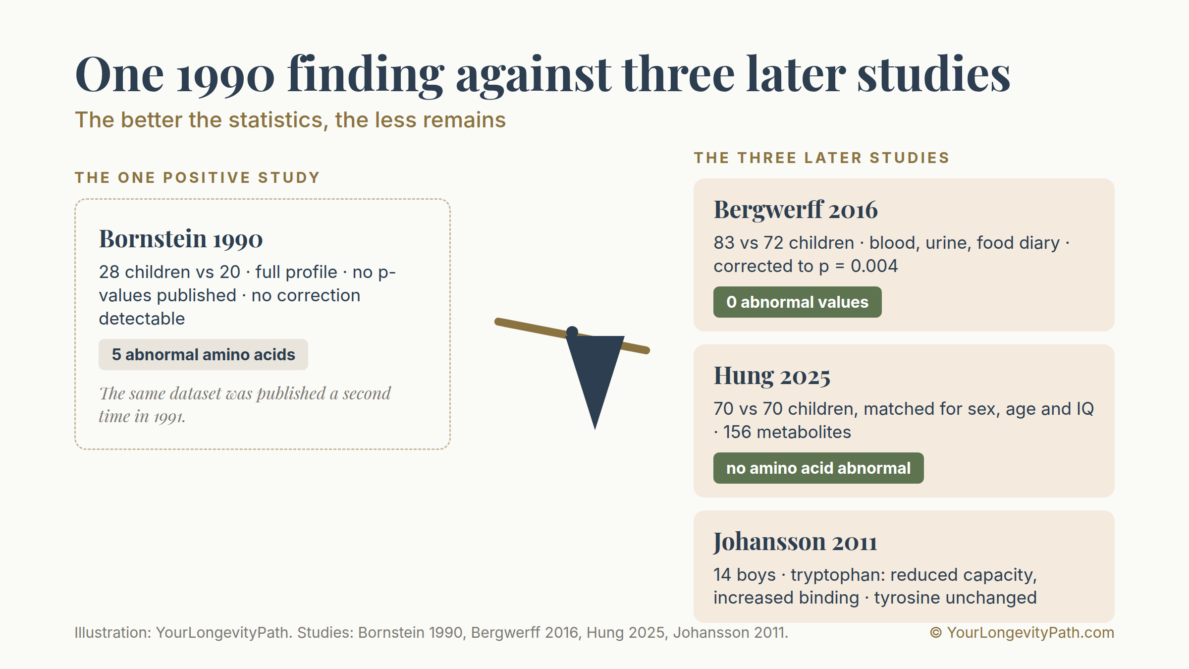
The narrative goes back to a single piece of work. In 1990 Bornstein and colleagues examined the blood of 28 children with attention disorder against 20 comparison children and found five amino acids lowered: phenylalanine, tyrosine, tryptophan, histidine and isoleucine. Their own interpretation in the abstract: a general deficiency in the uptake or transport of amino acids (Bornstein et al., 1990).
Two things about this before the finding travels on. Apart from the case numbers, the abstract contains not a single figure, no p-value, no effect size and no statement of which amino acids were unchanged. And the second paper from the same group, often cited as confirmation, examined an overlapping sample (Baker et al., 1991). Bergwerff and colleagues explicitly describe the two publications as two papers that describe the same sample (Bergwerff et al., 2016). It is one finding that was published twice.
Against this stand three newer studies:
Bergwerff 2016 examined 155 children between 6 and 13 years old, 83 of them with ADHD. Measurement was in dried blood, additionally in 18-hour urine, and protein intake was recorded with a food diary. The result: normal levels of the aromatic amino acids in blood and urine and a normal protein intake compared with the control group. The odds ratios for below-average values all went in the same direction, none reached significance: phenylalanine 2.22 (p = 0.07), tryptophan 2.09 (p = 0.11), tyrosine 1.83 (p = 0.22).
Hung 2025 compared 70 children who had never received an ADHD medication with 70 children matched for sex, age and IQ. 156 metabolites were measured. Four of them stood out: cholic acid, homoveratric acid, inosine and nicotinuric acid. Not a single amino acid.
Johansson 2011 examined transport in connective-tissue cells from 14 boys with ADHD. For tryptophan the transport capacity was reduced, the binding affinity by contrast increased, and for alanine the capacity was greater than in the controls. Tyrosine did not differ.
So a finding from 1990 stands against three later studies, among them the one with the best comparison method and the only one that corrected statistically for the number of measured values.
Why the studies report many hits and still show nothing
Here lies the real reason the field looks so contradictory, and it has nothing to do with ADHD.
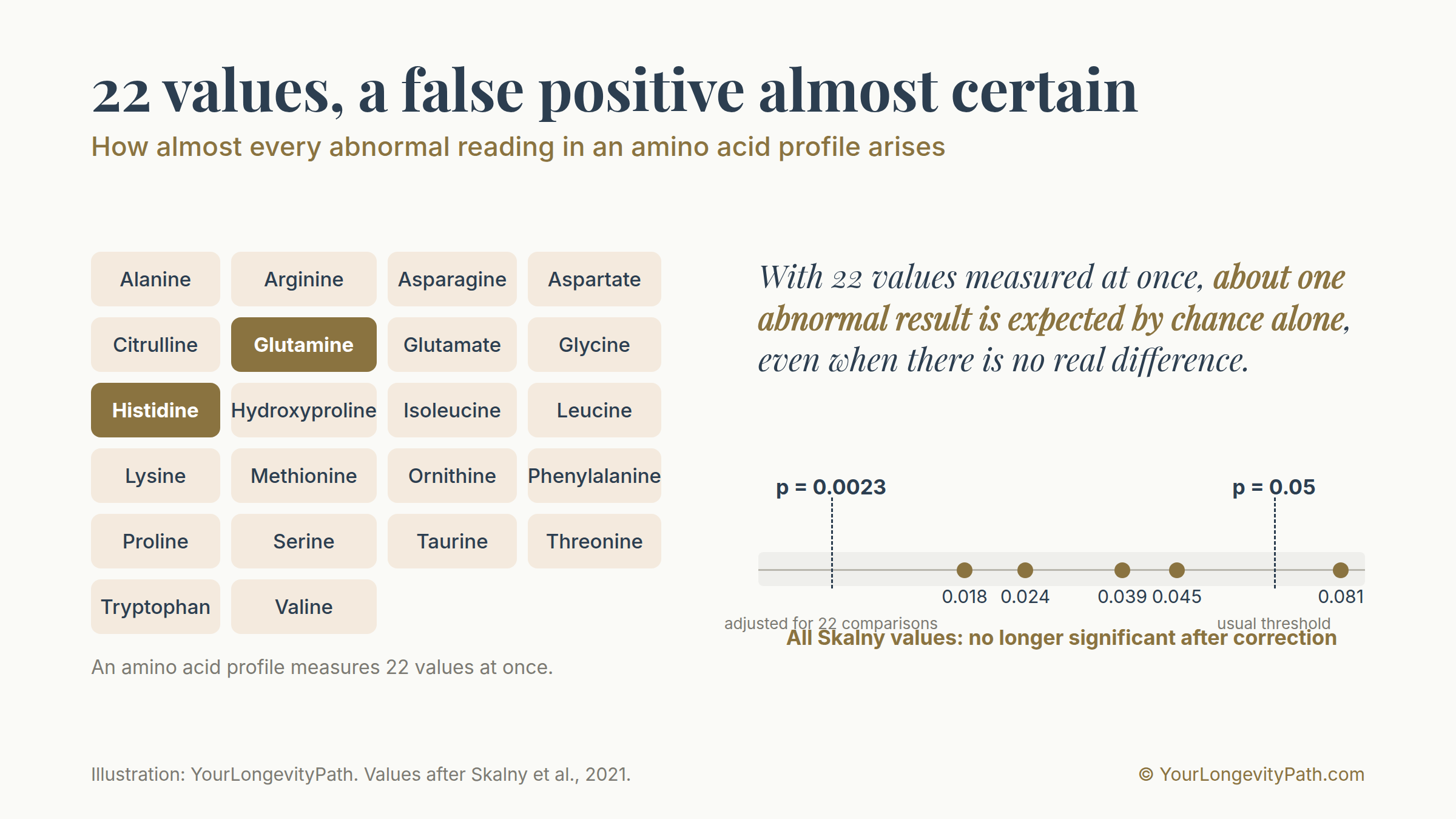
A plasma amino acid profile measures 20 to 22 values at once. At the usual significance threshold of 5 per cent, about one abnormal result per study is expected by chance alone, even when there is no difference between the groups. Anyone who tosses 22 coins inevitably gets a few striking runs. The only thing that helps against this is to adjust the threshold to the number of comparisons.
| Study | values measured | hits reported | p-values | correction |
|---|---|---|---|---|
| Bornstein 1990 | full profile | 5 | not reported | none detectable |
| Skalny 2021 | 22 | 4 to 5 | 0.018 to 0.045 | only within the ANCOVA, not across the 22 values |
| Johansson 2011 | several | 3 | 0.010 to 0.039 | none detectable |
| Bergwerff 2016 | 3 aromatic | 0 | all above 0.05 | yes, threshold 0.004 |
With Skalny’s 22 values, the adjusted threshold would be p = 0.0023. The lowest value reported there is 0.018, about eight times above it.
Of all the profile studies, exactly one corrected across all measured amino acids. It is at the same time the only one with a null result. There is a simple explanation for this: once you calculate correctly, nothing remains.
What happens when you look in adults
And here comes the point that is probably the most important for you. The entire profile literature on ADHD is a children’s literature. A complete plasma amino acid profile in adults with ADHD does not exist. Bornstein, Bergwerff, Skalny, Hung, Johansson: all children between 6 and 14 years old.
That does not mean nothing exists on adults. Individual metabolic pathways have certainly been measured there. What is missing is the measurement that would answer your question, namely the complete profile compared with a control group.
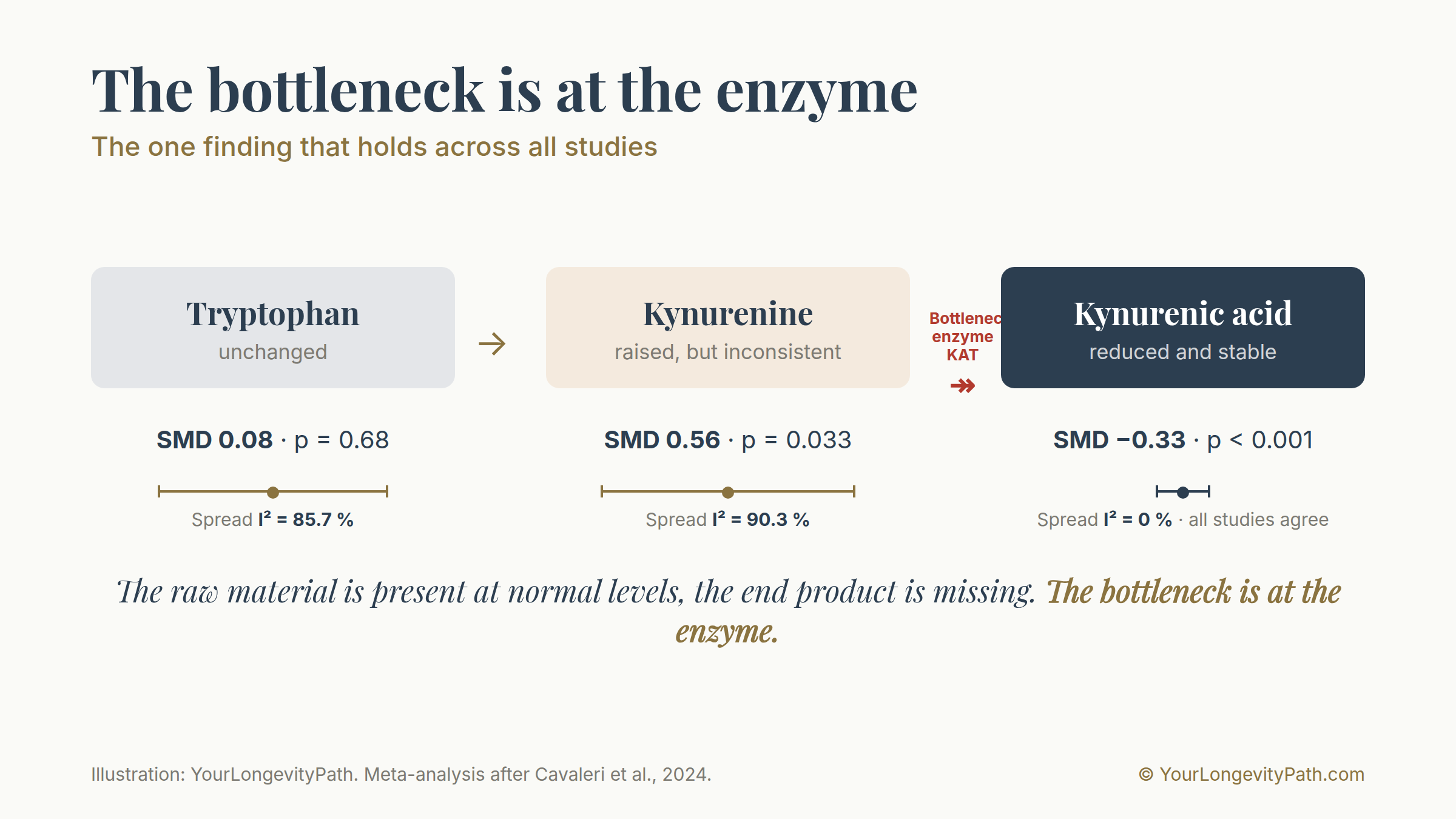
And where adults have been examined, something different shows up than in children. The meta-analysis of tryptophan metabolism evaluated 8 studies with 1,177 participants. In the overall analysis, tryptophan does not differ between ADHD and controls (SMD 0.08; p = 0.68). The variation between studies, however, is enormous: I², the measure of how strongly the individual studies diverge, is 85.7 per cent, where 0 per cent means complete agreement and 100 per cent maximum inconsistency. In the subgroup of medication-free children, tryptophan is then raised (SMD 0.31; 95% CI 0.11 to 0.50; p = 0.002), and this finding is statistically clean with I² = 0 per cent (Cavaleri et al., 2024). In adults the depletion studies point the other way (Zimmermann et al., 2012).
This reversal of direction is one reason not to transfer the children’s results to adults. When the same quantity comes out in opposite directions in two age groups, the average over both says nothing about either one.
For you as an adult with ADHD this means two things. The question is legitimate, and the existing studies answer it for eight-year-olds.
Amino acid by amino acid
Tyrosine. Not reproducibly lowered, and that is remarkable, because the entire dopamine-precursor narrative rests on tyrosine. One finding from 1990 against three negative studies. Skalny did not find tyrosine among the changed values, Hung did not find it among 156 metabolites.
Tryptophan. No deficit in the meta-analysis (p = 0.68), raised in medication-free children. The causal test has also been done: 14 studies specifically depleted tryptophan. If a tryptophan deficiency were a driver of ADHD symptoms, they would have to worsen during this. Only two of these works examined attention at all, with mixed results, and not a single one measured hyperactivity (Dinu et al., 2022). Controlled studies that give tryptophan in ADHD do not exist.
Histidine. The most-cited single value in the whole field, and the most important correction of this research. Skalny reports a serum value 29 per cent lower in children with ADHD. The associated p-value stands at 0.081. The study itself defines 0.05 as the threshold in its methods section. The difference is therefore not significant there, and the 29 per cent stand in the abstract without a p-value. There is no third histidine study, no intervention studies either, and the only measurement of histamine in ADHD so far found no association with inattention or hyperactivity (Bruton et al., 2024).
Glutamine and glutamate. Contradictory, and the methodologically strongest works are negative. Apart from Skalny (p = 0.024, uncorrected across 22 tests), glutamine was not measured in any further dedicated blood study. For glutamate, the largest study with 145 against 120 participants found no difference (p = 0.059; Cheng et al., 2026); Hung does not find it among his results.
GABA. The most solid blood finding in the whole field, and it argues against supplements. In serum, GABA is raised in ADHD, with p < 0.001 and a discriminatory power of 0.87; it correlates with symptom severity and falls under methylphenidate (Cheng et al., 2026; Miniksar et al., 2022). In the brain the finding is unremarkable in meta-analysis (Schür et al., 2016). On top of this comes the blood-brain barrier: it actively transports GABA outward, and the efflux clearance is at least 16 times higher than the influx clearance (Kakee et al., 2001). A value that is raised in the blood and rises with symptom severity, plus a barrier that actively pumps it out, is no argument for GABA capsules. Anyone wanting to work on the excitation-inhibition balance finds the better-supported point at magnesium, which blocks the NMDA receptor: magnesium in ADHD.
Taurine. The biggest nothing in the field. There is not a single study that has measured taurine in people with ADHD and compared it with a control group, and none that has tested it as a treatment. Skalny measured taurine along the way, it is in the list of 22 values, and it is not among the changed ones. A large part of the “taurine improves attention” literature comes from energy-drink studies in which taurine was given together with caffeine and sugar. There the effect cannot be attributed to the taurine.
Protein intake. Two independent studies with direct dietary assessment find no difference (Bergwerff et al., 2016; Holton et al., 2019). The often-told chain from low protein intake via low tyrosine to low dopamine is already unsupported at its first step.
The one finding that holds
For all the criticism, something remains, and it is more interesting than what is usually told.
Of all the values in tryptophan metabolism, exactly one is stable across the studies: kynurenic acid is lowered (SMD −0.33; 95% CI −0.49 to −0.17; p < 0.001), and with a variation between studies of I² = 0 per cent. For comparison: for the raised kynurenine, which is cited more often, the same figure is 90.3 per cent, so the studies there contradict each other considerably (Cavaleri et al., 2024). The finding was replicated in children who had never received an ADHD medication, and there tryptophan, kynurenine and the ratio of the two were all unchanged (Ünal et al., 2026).
Kynurenic acid is formed from kynurenine via a particular enzyme. When the raw material is present at normal levels and the end product is missing, the narrow point lies at this enzyme. That is a different statement from “tryptophan is missing”, and it cannot be captured by an amino acid profile.
One limitation belongs here. The study most often cited for a shift in metabolism in adults measured eight values at once and did not correct. There kynurenine itself was not significant (p = 0.15), and after accounting for the tryptophan level only three values remained. On top of this comes a comparison problem: 66.2 per cent of the patient group were smokers by nicotine level against 12.2 per cent of the control group, and smoking lowers tryptophan (Aarsland et al., 2015).
Would more of it even help?
Suppose a building block really were missing. Then “more of it” would still not be the answer, and this is well studied in healthy adults.
The review on tyrosine summarises the conditions like this: it works only when the transmitter system is intact and dopamine or noradrenaline are temporarily reduced (Jongkees et al., 2015). Two prerequisites at the same time, and the evidence for them comes from cold exposure, from staying awake for over 24 hours, and from a military training course.
The opposite direction is the more revealing part:
- In a double-blind study with 45 people and 2 grams of tyrosine, the weaker participants improved, the stronger ones got worse. The group average did not change, the effect showed only in the spread (Jongkees, 2020).
- In 70 people, tyrosine worsened mental flexibility under high cognitive load. The authors call this themselves the opposite of what they had expected (Robson et al., 2020).
- In older adults, working memory worsened in a dose-dependent way, most clearly in those with the highest rise in the blood (van de Rest et al., 2017).
Tyrosine therefore shifts the position on a curve with an optimum. Anyone already well placed slides past it.
The only tyrosine study in ADHD shows the same pattern over time. Twelve adults received L-tyrosine for eight weeks; eight of them improved after two weeks, and after six weeks the effect had disappeared. The authors write that L-tyrosine is without benefit in attention disorder (Reimherr et al., 1987). In supplement marketing this regularly becomes “8 of 12 responded”. A randomised controlled trial of L-tyrosine in ADHD does not exist to this day.
One note belongs here. The most frequently cited work for amino acid precursors in ADHD, reporting 77 per cent improvement, was retracted in 2020. It was a retrospective chart review, and the first author himself sells corresponding supplements (Hinz et al., 2011, retracted 2020).
Taurine, glycine, tyrosine: what is proven for the supplements
Up to here it has been about status, that is, the question of whether something is missing. The second question is a different one: does one of these amino acids work as a supplement, regardless of whether a deficiency is present? Does taurine calm, does tyrosine make you briefly more alert, does glycine relax? This question is better studied than the first, and the answers are more concrete.
One pattern runs through all three. Taking it away is proven in humans, adding it is not. For tyrosine there is a study that measured in the brain scanner what a tyrosine depletion does: the amphetamine-triggered dopamine release in the striatum falls in eight men, in a linear relationship with the drop in the blood (r = −0.82; Leyton et al., 2004). The same shows via the prolactin level (Harmer et al., 2001). A study that measures a rise in dopamine release in humans after giving tyrosine does not exist.
From “taking it away lowers” it does not follow, for this enzyme, that “adding it raises”. Tyrosine hydroxylase is braked by its own end product; dopamine binds with high affinity and blocks it until the neuron fires and the brake is released (Gordon et al., 2008). The specialists from whom the whole precursor narrative originates limit their own finding accordingly: more tyrosine increases catecholamine production only in nerve cells that are currently active (Fernstrom & Fernstrom, 2007).
On top of this comes a curve that explains the rest. In animal experiments, tyrosine levels and synthesis rate in the brain rise as the dietary protein content increases from 2 to 10 per cent. Between 10 and 20 per cent, nothing more happens (Fernstrom & Fernstrom, 1995). The synthesis rate rose only in the retina and hypothalamus, not in the prefrontal cortex, that is, precisely not in the region that matters in ADHD. Anyone eating normally is already sitting on this plateau.
Taurine: the blood-brain barrier is the point, not the study record.
The transporter that brings taurine into the brain has been measured in a human model. It runs half-saturated at about 19 micromoles per litre (Tega et al., 2025). After 4 grams of taurine, the peak level in the blood is around 690 micromoles per litre (Ghandforoush-Sattari et al., 2010). The transporter is therefore occupied far beyond its saturation point and already runs near its maximum at normal levels. In addition there is an active pump that carries taurine out of the brain (Lee & Kang, 2004), and when taurine is in excess the transporter is turned down at the level of gene expression (Kang et al., 2002). The half-life in the blood is about one hour.
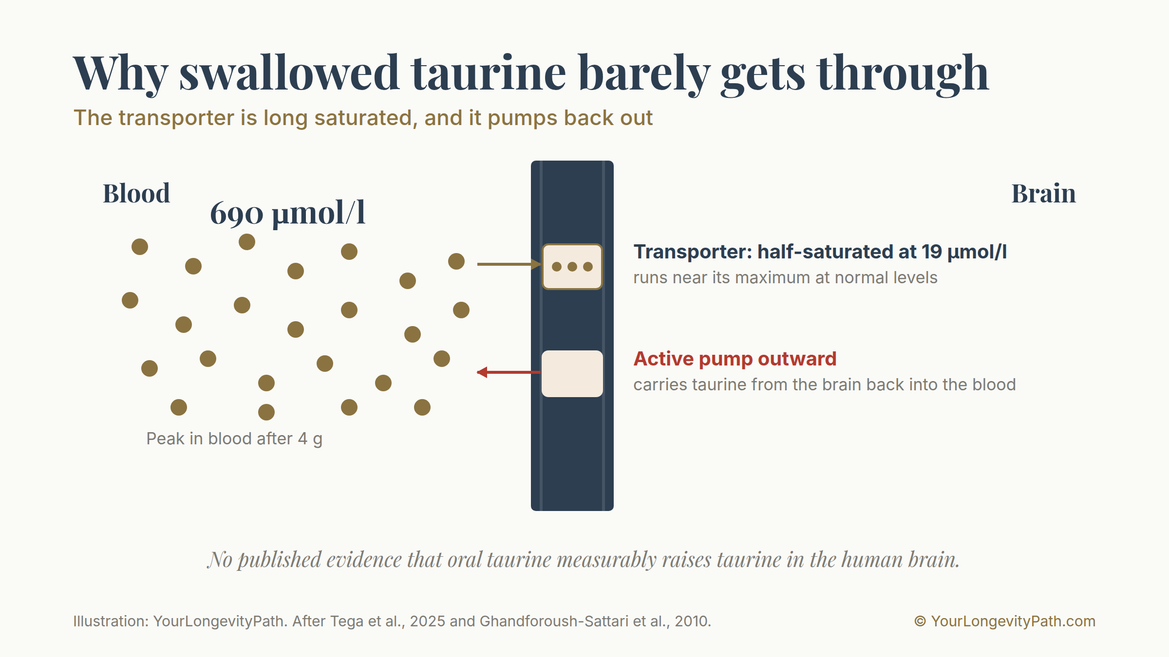
There is no published evidence anywhere that oral taurine measurably raises taurine in the human brain, neither via magnetic resonance spectroscopy nor via the cerebrospinal fluid level. And there is a direct counter-indication: in one patient group, taurine was raised in the blood and unchanged in the cerebrospinal fluid (Samuelsson et al., 2013).
The study record fits this. A review of all eight randomised studies on taurine and cognition in humans concludes that acute doses produced at best small and inconsistent improvements and that most endpoints remained unchanged (Moore et al., 2026). The meta-analysis of 7 studies with 402 people finds no effect (Cao et al., 2025). On sleep there is not a single randomised study, on ADHD not a single intervention study. The review on mood counts one human study against 13 animal studies, and the one human study is an observational study with no association (Pedregosa et al., 2026).
And even in the animal model the finding does not hold reliably. One work that tested this specifically found no effect on anxiety- or depression-like behaviour and no stimulating effect (Whirley & Einat, 2008).
Where the calming claim then comes from can be traced back. It begins with a correct finding. Taurine does indeed act at the glycine and the GABA-A receptor; that has been measured since the eighties. The half-maximal effective concentration at the glycine receptor is 406 micromoles per litre (Hussy et al., 1997), at some preparations 1,250 (Jiang et al., 2004). Taurine is moreover only a partial agonist there, reaching at most 70 per cent of the maximal glycine response (Hussy et al., 1997). In the brain, the actual concentration between the cells is 25 micromoles per litre, measured in the rat striatum (Molchanova et al., 2004). The receptor experiments therefore run far above anything that ever occurs in the head.
The second step is the route of administration. The animal works that show a dampening effect gave taurine directly into the brain. And one work compared both routes in the same model. The result: injected into the brain, taurine lengthened sleep; swallowed, it shortened it (McBroom et al., 1986). The authors name the reason themselves, namely the slow uptake into the brain. At the same time, other groups were building fat-soluble taurine derivatives, expressly because it was assumed they would cross the blood-brain barrier (Ahtee et al., 1985). The problem was known in 1985.
The third finding goes beyond the mere absence of an effect. In mice, taurine injected once is anxiolytic, given continuously in the drinking water by contrast anxiogenic (El Idrissi et al., 2009). The reason lies in adaptation: under constant taurine supply, the GABA-A receptors are downregulated (L’Amoreaux et al., 2010). A regulated system evades the constant stimulus.
Two things round this off. Oral taurine has long been tested in humans at an endpoint of brain excitability: in the seventies a series of epilepsy studies ran with up to 8 grams daily. The last of them gave six patients 375 to 8,000 milligrams per day and found no improvement in seizure control (Mantovani & DeVivo, 1979). This literature is 45 years old and is cited nowhere. And with the European food authority, six health claims were submitted for taurine for assessment, among them immune system, heart, muscle and cognition. Not a single one on relaxation, anxiety or sleep (EFSA, 2011). The effect for which taurine is sold today was never submitted for assessment.
For accuracy’s sake, two transport studies contradict each other on whether the active transporter sits on both sides of the vessel wall or only on the side facing the brain (Tamai et al., 1995; Rasgado-Flores et al., 2012). This changes nothing about the overall picture.
Two things belong to accuracy. First, the only place where taurine does something calming-like in humans is the comparison with caffeine: in the two studies that tested taurine in isolation, it dampens the caffeine effect rather than stimulating on its own (Giles et al., 2012; Peacock et al., 2013). Both works attribute the entire energy-drink effect to the caffeine, and neither measured anxiety. Second, there is one positive study: 4 grams of taurine over twelve weeks improved the symptoms in first-episode psychosis (p = 0.004). That comes with 29 per cent dropouts, a second primary endpoint on cognition with no effect (p = 0.582) and a population that has nothing to do with this question (O’Donnell et al., 2016).
Glycine: the source record is the problem.
The two studies on which the story of sleep-promoting glycine rests are not findable in the medical databases (Yamadera et al., 2007; Inagawa et al., 2006). No abstract, no p-values, no conflict-of-interest statement. The participant numbers were 11 and 19.
All primary works come from a single company, the largest amino acid manufacturer in the world. The only fully verifiable of these studies has seven evaluated participants, is only single-blind, was approved by the company’s own ethics committee and states in the text that there is no commercial relationship (Bannai et al., 2012). The best available review summarises the sleep evidence like this: small samples with a high risk of bias, and a meta-analysis was not possible because of the inconsistency (Soh et al., 2024).
One work with real laboratory sleep measurement tested glycine-rich collagen peptides in 13 men. Sleep onset, sleep amount, sleep efficiency and core body temperature remained unchanged; the number of waking events fell. This work, too, was carried out with a manufacturer’s involvement (Thomas et al., 2024).
The finding that settles the matter, however, is a different one. A double-blind study gave glycine by vein, 7 to 14 grams, and afterwards measured the cerebrospinal fluid. The level there rose dose-dependently without producing any clear effect in the central nervous system (D’Souza et al., 2000). It arrived in the brain and did nothing measurable there. If the multiple dose is ineffective even when bypassing the digestive tract, a noticeable effect from 3 grams out of a can is hard to justify.
The outcome of the large clinical trial fits with this. In schizophrenia research, glycine was tested in high doses; small studies had reported improvements of 23 to 30 per cent. The adequately powered study with 157 participants found no difference from placebo either in symptoms or in cognition, and this under the co-authorship of the very researchers who had previously published the positive small studies (Buchanan et al., 2007).
The time axis is the part almost no one mentions.
In ADHD, amino acids were given over a longer period twice, and both times the same thing happened. Under L-tyrosine, eight of twelve adults improved after two weeks; after six weeks the effect was gone (Reimherr et al., 1987). Under DL-phenylalanine, a double-blind effect on mood appeared, and in the subsequent open continuation the benefit disappeared completely within three months (Wood et al., 1985). Two different amino acids, the same pattern: an initial effect, then regulation catches up with it.
The most recent work on this is a randomised controlled trial of 5-HTP, the direct precursor of serotonin, in 112 adults with pronounced ADHD traits. Result: no benefit on any measure of distractibility, the only significant result ran in the wrong direction, and 19.6 per cent had side effects with fatigue, nausea or vomiting (Jackson et al., 2026).
| Randomised trial with a fitting endpoint | Evidence in the human brain | Tested in ADHD | |
|---|---|---|---|
| Tyrosine | only under acute stress or exhaustion | only for depletion | open trial, effect lost after 6 weeks |
| Taurine | none on anxiety or sleep | none | none |
| Glycine | three small ones, all from the manufacturer | rise without effect | none |
What this does not mean. A lack of evidence is not evidence of ineffectiveness. Taurine research in humans consists almost entirely of single doses in healthy people; longer administration with psychological endpoints is practically untested. At 3 grams daily, taurine is well tolerated, and a self-experiment is defensible. What cannot be derived from these data is a promise of effect.
Does a stool test show whether I absorb protein poorly?
This question is legitimate, because the one positive amino acid study points there itself. Bornstein interpreted his values as an uptake or transport disorder. The answer consists of two parts.
First, the hypothesis has never been tested. No study in ADHD has measured pancreatic elastase in stool, protein loss through the gut, faecal nitrogen or an absorption test with labelled protein. In ADHD, the only things ever examined in stool at all are the microbiome, once the fungal flora and once calprotectin, the latter with no difference in 14 children (Soltysova et al., 2025). That is a real research gap.
Second, a stool test would be the wrong tool for this. A study in 11 patients with confirmed pancreatic insufficiency shows this directly. With labelled protein the absorption disorder could be measured and an improvement demonstrated under enzyme therapy. In the stool none of this was visible: a protein absorption disorder was largely undetectable with the usual stool tests (Airinei et al., 2011).
The reason for this is vivid. The nitrogen in stool comes mainly from bacterial mass, shed intestinal cells and digestive secretions. What the small intestine does not absorb is used up by the large-intestine bacteria before it reaches the stool.
On the permeability values that such profiles come with, a note of its own belongs. The best known of them has been checked several times, and according to two independent studies the test kits used detect proteins other than what is stated on the report (Scheffler et al., 2018; Ajamian et al., 2019). The two larger and better-adjusted studies in ADHD find no difference there anyway (Aydoğan Avşar et al., 2021; Lee et al., 2023).
And what if IBS is also present?
Irritable bowel syndrome is indeed more common in ADHD. An analysis across 11 studies and around 3.85 million people found no increased value for intestinal disorders overall (OR 1.25; 95% CI 0.75 to 2.07), but did for irritable bowel syndrome (OR 1.63; 1.45 to 1.83; Ng et al., 2025). A large cohort confirms this and adds two things: inflammatory bowel disease and coeliac disease are not increased in ADHD, and methylphenidate itself increases digestive complaints and constipation (Kedem et al., 2020). This fits the mechanism: as a stimulant it dampens appetite and digestive activity, the gut becomes sluggish, and often less is drunk as well.
The obvious conclusion would now be that in IBS absorption suffers and from this an amino acid deficiency arises. This chain breaks in the middle. An absorption disorder is an exclusion criterion in IBS: it is the diagnosis you give after malabsorption has been ruled out. Empirically this fits. In an analysis of over 12,000 people, those with IBS actually consumed more calories than healthy people, with no signs of nutrient deficiencies (Hujoel et al., 2020). Where deficiencies are found, the author groups attribute them to reduced intake, that is, to avoidance behaviour and elimination diets.
In two places, however, the objection is right after all:
Missed coeliac disease. In an analysis of 29 studies with 7,209 people with an IBS diagnosis, 2 per cent had biopsy-confirmed coeliac disease, 6 per cent had abnormal antibodies, and 15 per cent of the abnormal ones were never investigated further (Shiha et al., 2025). Coeliac disease damages the small-intestine surface and causes a genuine protein malabsorption.
Missed pancreatic insufficiency. In the diarrhoea form of IBS, stool elastase is below normal in 6.1 per cent of those affected, against 0 per cent in two control groups (Leeds et al., 2010). The pancreas delivers the protein-splitting enzymes; it is the only one of the mentioned mechanisms that acts directly on protein digestion.
In both cases the chain holds completely. But then there is a missed diagnosis and not IBS. From this comes a usable rule. If someone with ADHD and an IBS diagnosis has measurable nutrient deficiencies, the next two questions are: what does this person actually eat, and has coeliac serology ever been done?
What you can do instead
1. An amino acid profile does not answer the ADHD question. Two questions are often mixed here. One is: in people with ADHD, is a particular amino acid typically lowered? A profile gives no answer to this, because for adults with ADHD there is no published comparison group. The other is: in you specifically, is something below the reference range? A profile does measure that, and that is what I use it for, together with the protein supply and the ratio of glutamine to glutamate.
A single abnormal value out of 22 measured, however, is not yet a finding. The way to tell them apart is the second measurement: what is low again on remeasuring was no coincidence.
2. Check the values for which there is good evidence. Ferritin, vitamin D, the omega-3 index, whole-blood magnesium and zinc are studied in ADHD, and for some of them there are intervention studies. Which values come in which order I have put together in ADHD and blood values.
3. Look at protein intake, entirely without a lab. On average it is unremarkable in ADHD, but individual people take in too little, especially in the morning. Anyone who notes it for a few days sees it quickly, and it costs nothing.
4. With persistent gut complaints, have it clarified specifically. If complaints persist, coeliac serology and, with diarrhoea, stool elastase belong in the picture, both through the doctor. In coaching we can, before and after, put into context which investigation is due and what the results mean for your everyday life.
5. Do not supplement without a reason. For tyrosine in ADHD there is no controlled study, for taurine not a single measurement, and for GABA the available data argue against it.
Don't get me wrong
No study evidence for an ADHD-typical amino acid deficiency does not mean you have no deficiency. It can exist, and targeted supplementation can help with symptoms. The point is a different one: an abnormal value on your report says something about you, not about ADHD. Separating the two is the real work, and supplementation belongs tailored to the individual case rather than to a diagnosis.
Where to go from here
The question of the building blocks is legitimate. Research’s answer at present is: measurement was done in children, the calculation was mostly wrong, and for adults the decisive study is missing. That is an unsatisfying state, and it is nonetheless usable, because it saves you expensive diagnostics and directs the view to the values where there is actually something to gain.
In my ADHD coaching for adults I look at your values together with your sleep, your diet and your everyday life, and sort out what comes first for you. You get your plan in writing, always within what coaching can do and what belongs in medical hands.
If you would first like to find out whether this fits you, book a free initial consultation. It is non-binding and takes place online.
Scientific sources
-
Aarsland, T. I., Landaas, E. T., Hegvik, T. A., et al. (2015). Serum concentrations of kynurenines in adult patients with attention-deficit hyperactivity disorder (ADHD): a case-control study. Behavioral and Brain Functions, 11, 36. doi: 10.1186/s12993-015-0080-x
-
Ahtee, L., Auvinen, H., Mäenpää, A. R., Vahala, M. L., Lehtinen, M., & Halmekoski, J. (1985). Comparison of central nervous system actions of taurine and N-pivaloyltaurine. Acta Pharmacologica et Toxicologica, 57(2), 96–105. doi: 10.1111/j.1600-0773.1985.tb00016.x
-
Airinei, G., Gaudichon, C., Bos, C., et al. (2011). Postprandial protein metabolism but not a fecal test reveals protein malabsorption in patients with pancreatic exocrine insufficiency. Clinical Nutrition, 30(6), 831–837. doi: 10.1016/j.clnu.2011.06.006
-
Ajamian, M., Steer, D., Rosella, G., & Gibson, P. R. (2019). Serum zonulin as a marker of intestinal mucosal barrier function: may not be what it seems. PLOS ONE, 14(1), e0210728. doi: 10.1371/journal.pone.0210728
-
Aydoğan Avşar, P., Işık, Ü., Aktepe, E., et al. (2021). Serum zonulin and claudin-5 levels in children with attention-deficit/hyperactivity disorder. International Journal of Psychiatry in Clinical Practice, 25(1), 49–55. doi: 10.1080/13651501.2020.1801754
-
Baker, G. B., Bornstein, R. A., Rouget, A. C., Ashton, S. E., van Muyden, J. C., & Coutts, R. T. (1991). Phenylethylaminergic mechanisms in attention-deficit disorder. Biological Psychiatry, 29(1), 15–22. doi: 10.1016/0006-3223(91)90207-3
-
Bannai, M., Kawai, N., Ono, K., Nakahara, K., & Murakami, N. (2012). The effects of glycine on subjective daytime performance in partially sleep-restricted healthy volunteers. Frontiers in Neurology, 3, 61. doi: 10.3389/fneur.2012.00061
-
Bergwerff, C. E., Luman, M., Blom, H. J., & Oosterlaan, J. (2016). No tryptophan, tyrosine and phenylalanine abnormalities in children with attention-deficit/hyperactivity disorder. PLOS ONE, 11(3), e0151100. doi: 10.1371/journal.pone.0151100
-
Bornstein, R. A., Baker, G. B., Carroll, A., et al. (1990). Plasma amino acids in attention deficit disorder. Psychiatry Research, 33(3), 301–306. doi: 10.1016/0165-1781(90)90046-8
-
Bruton, A. M., Robinette, L. M., Hatsu, I. E., et al. (2024). Urinary histamine not associated with severity of symptoms of ADHD and emotional dysregulation: a cross-sectional secondary data analysis from the Micronutrients for ADHD in Youth (MADDY) study. Advances in Neurodevelopmental Disorders, 9, 595–606. doi: 10.1007/s41252-024-00432-y
-
Buchanan, R. W., Javitt, D. C., Marder, S. R., et al. (2007). The Cognitive and Negative Symptoms in Schizophrenia Trial (CONSIST): the efficacy of glutamatergic agents for negative symptoms and cognitive impairments. American Journal of Psychiatry, 164(10), 1593–1602. doi: 10.1176/appi.ajp.2007.06081358
-
Cao, Q., Nie, Z., Liu, Y., Xu, J., Chen, L., & Han, S. (2025). Effects of taurine supplementation on cognitive function: a systematic review and meta-analysis of randomised controlled trials. International Journal of Food Sciences and Nutrition, 76(4), 370–380. doi: 10.1080/09637486.2025.2499044
-
Cavaleri, D., Crocamo, C., Morello, P., Bartoli, F., & Carrà, G. (2024). The kynurenine pathway in attention-deficit/hyperactivity disorder: a systematic review and meta-analysis of blood concentrations of tryptophan and its catabolites. Journal of Clinical Medicine, 13(2), 583. doi: 10.3390/jcm13020583
-
Cheng, X., Wang, L., Diao, C., Wang, Y., & Xue, Y. (2026). The ratio of GABA/Glu as a biomarker in children with attention deficit hyperactivity disorder. Physiology & Behavior, 315, 115445. doi: 10.1016/j.physbeh.2026.115445
-
D’Souza, D. C., Gil, R., Cassello, K., et al. (2000). IV glycine and oral D-cycloserine effects on plasma and CSF amino acids in healthy humans. Biological Psychiatry, 47(5), 450–462. doi: 10.1016/S0006-3223(99)00133-X
-
Dinu, L. M., Phattharakulnij, N., & Dommett, E. J. (2022). Tryptophan modulation in individuals with attention deficit hyperactivity disorder: a systematic review. Journal of Neural Transmission, 129(4), 361–377. doi: 10.1007/s00702-022-02478-5
-
EFSA Panel on Dietetic Products, Nutrition and Allergies (2011). Scientific opinion on the substantiation of health claims related to taurine. EFSA Journal, 9(4), 2035. doi: 10.2903/j.efsa.2011.2035
-
El Idrissi, A., Boukarrou, L., Heany, W., Malliaros, G., Sangdee, C., & Neuwirth, L. (2009). Effects of taurine on anxiety-like and locomotor behavior of mice. Advances in Experimental Medicine and Biology, 643, 207–215. doi: 10.1007/978-0-387-75681-3_21
-
Fernstrom, M. H., & Fernstrom, J. D. (1995). Effect of chronic protein ingestion on rat central nervous system tyrosine levels and in vivo tyrosine hydroxylation rate. Brain Research, 672(1–2), 97–103. doi: 10.1016/0006-8993(94)01342-F
-
Fernstrom, J. D., & Fernstrom, M. H. (2007). Tyrosine, phenylalanine, and catecholamine synthesis and function in the brain. Journal of Nutrition, 137(6), 1539S–1547S. doi: 10.1093/jn/137.6.1539S
-
Ghandforoush-Sattari, M., Mashayekhi, S., Krishna, C. V., Thompson, J. P., & Routledge, P. A. (2010). Pharmacokinetics of oral taurine in healthy volunteers. Journal of Amino Acids, 2010, 346237. doi: 10.4061/2010/346237
-
Giles, G. E., Mahoney, C. R., Brunyé, T. T., Gardony, A. L., Taylor, H. A., & Kanarek, R. B. (2012). Differential cognitive effects of energy drink ingredients: caffeine, taurine, and glucose. Pharmacology, Biochemistry, and Behavior, 102(4), 569–577. doi: 10.1016/j.pbb.2012.07.004
-
Gordon, S. L., Quinsey, N. S., Dunkley, P. R., & Dickson, P. W. (2008). Tyrosine hydroxylase activity is regulated by two distinct dopamine-binding sites. Journal of Neurochemistry, 106(4), 1614–1623. doi: 10.1111/j.1471-4159.2008.05509.x
-
Harmer, C. J., McTavish, S. F., Clark, L., Goodwin, G. M., & Cowen, P. J. (2001). Tyrosine depletion attenuates dopamine function in healthy volunteers. Psychopharmacology, 154(1), 105–111. doi: 10.1007/s002130000613
-
Hinz, M., Stein, A., Neff, R., Weinberg, R., & Uncini, T. (2011). Treatment of attention deficit hyperactivity disorder with monoamine amino acid precursors and organic cation transporter assay interpretation. Neuropsychiatric Disease and Treatment, 7, 31–38. Retracted 2020: Neuropsychiatric Disease and Treatment, 16, 3131. doi: 10.2147/NDT.S16270
-
Holton, K. F., Johnstone, J. M., Brandley, E. T., & Nigg, J. T. (2019). Evaluation of dietary intake in children and college students with and without attention-deficit/hyperactivity disorder. Nutritional Neuroscience, 22(9), 664–677. doi: 10.1080/1028415X.2018.1427661
-
Hujoel, I. A. (2020). Nutritional status in irritable bowel syndrome: a North American population-based study. JGH Open, 4(4), 656–662. [PMID: 32782953] doi: 10.1002/jgh3.12311
-
Hung, Y. A., Kuo, T. C., Tseng, Y. J., Shang, C. Y., & Gau, S. S. (2025). Identifying novel metabolites in children with attention-deficit/hyperactivity disorder through metabolome profiling. Translational Psychiatry, 15(1), 180. doi: 10.1038/s41398-025-03393-5
-
Hussy, N., Deleuze, C., Pantaloni, A., Desarménien, M. G., & Moos, F. (1997). Agonist action of taurine on glycine receptors in rat supraoptic magnocellular neurones: possible role in osmoregulation. Journal of Physiology, 502(3), 609–621. doi: 10.1111/j.1469-7793.1997.609bj.x
-
Inagawa, K., Hiraoka, T., Kohda, T., Yamadera, W., & Takahashi, M. (2006). Subjective effects of glycine ingestion before bedtime on sleep quality. Sleep and Biological Rhythms, 4(1), 75–77. doi: 10.1111/j.1479-8425.2006.00193.x
-
Jackson, E., Riley, T., & Overton, P. G. (2026). The effect of 5-hydroxytryptophan, a serotonin precursor, on adults with high levels of attention deficit hyperactivity disorder traits: a randomised, controlled trial. PLOS ONE, 21(5), e0349512. doi: 10.1371/journal.pone.0349512
-
Jiang, Z., Krnjević, K., Wang, F., & Ye, J. H. (2004). Taurine activates strychnine-sensitive glycine receptors in neurons freshly isolated from nucleus accumbens of young rats. Journal of Neurophysiology, 91(1), 248–257. doi: 10.1152/jn.00106.2003
-
Johansson, J., Landgren, M., Fernell, E., et al. (2011). Altered tryptophan and alanine transport in fibroblasts from boys with attention-deficit/hyperactivity disorder (ADHD): an in vitro study. Behavioral and Brain Functions, 7, 40. doi: 10.1186/1744-9081-7-40
-
Jongkees, B. J., Hommel, B., Kühn, S., & Colzato, L. S. (2015). Effect of tyrosine supplementation on clinical and healthy populations under stress or cognitive demands: a review. Journal of Psychiatric Research, 70, 50–57. doi: 10.1016/j.jpsychires.2015.08.014
-
Jongkees, B. J. (2020). Baseline-dependent effect of dopamine’s precursor L-tyrosine on working memory gating but not updating. Cognitive, Affective & Behavioral Neuroscience, 20(3), 521–535. doi: 10.3758/s13415-020-00783-8
-
Kakee, A., Takanaga, H., Terasaki, T., Naito, M., Tsuruo, T., & Sugiyama, Y. (2001). Efflux of a suppressive neurotransmitter, GABA, across the blood-brain barrier. Journal of Neurochemistry, 79(1), 110–118. [PMID: 11595763] doi: 10.1046/j.1471-4159.2001.00540.x
-
Kang, Y. S., Ohtsuki, S., Takanaga, H., Tomi, M., Hosoya, K., & Terasaki, T. (2002). Regulation of taurine transport at the blood-brain barrier by tumor necrosis factor-alpha, taurine and hypertonicity. Journal of Neurochemistry, 83(5), 1188–1195. doi: 10.1046/j.1471-4159.2002.01223.x
-
Kedem, S., Yust-Katz, S., Carter, D., et al. (2020). Attention deficit hyperactivity disorder and gastrointestinal morbidity in a large cohort of young adults. World Journal of Gastroenterology, 26(42), 6626–6637. doi: 10.3748/wjg.v26.i42.6626
-
L’Amoreaux, W. J., Marsillo, A., & El Idrissi, A. (2010). Pharmacological characterization of GABA-A receptors in taurine-fed mice. Journal of Biomedical Science, 17(Suppl 1), S14. doi: 10.1186/1423-0127-17-S1-S14
-
Lee, N. Y., & Kang, Y. S. (2004). The brain-to-blood efflux transport of taurine and changes in the blood-brain barrier transport system by tumor necrosis factor-alpha. Brain Research, 1023(1), 141–147. doi: 10.1016/j.brainres.2004.07.033
-
Lee, S. Y., Li, S. C., Yang, C. Y., Kuo, H. C., Chou, W. J., & Wang, L. J. (2023). Gut leakage markers and cognitive functions in patients with attention-deficit/hyperactivity disorder. Children, 10(3), 513. doi: 10.3390/children10030513
-
Leeds, J. S., Hopper, A. D., Sidhu, R., et al. (2010). Some patients with irritable bowel syndrome may have exocrine pancreatic insufficiency. Clinical Gastroenterology and Hepatology, 8(5), 433–438. doi: 10.1016/j.cgh.2009.09.032
-
Leyton, M., Dagher, A., Boileau, I., et al. (2004). Decreasing amphetamine-induced dopamine release by acute phenylalanine/tyrosine depletion: a PET/[11C]raclopride study in healthy men. Neuropsychopharmacology, 29(2), 427–432. doi: 10.1038/sj.npp.1300328
-
Mantovani, J., & DeVivo, D. C. (1979). Effects of taurine on seizures and growth hormone release in epileptic patients. Archives of Neurology, 36(11), 672–674. doi: 10.1001/archneur.1979.00500470042006
-
McBroom, M. J., Elkhawad, A. O., & Dlouha, H. (1986). Taurine and ethanol-induced sleeping time in mice: route and time course effects. General Pharmacology, 17(1), 97–100. doi: 10.1016/0306-3623(86)90018-2
-
Miniksar, D. Y., Cansız, M. A., Kılıç, M., & Göçmen, A. Y. (2022). Relationship between sleep problems and chronotypes of children and adolescents with attention deficit and hyperactivity disorder and serum GABA, glutamate and homocysteine levels. Chronobiology International, 39(3), 386–397. doi: 10.1080/07420528.2021.2018452
-
Molchanova, S., Oja, S. S., & Saransaari, P. (2004). Characteristics of basal taurine release in the rat striatum measured by microdialysis. Amino Acids, 27(3–4), 261–268. doi: 10.1007/s00726-004-0139-8
-
Moore, J. A., Cousins, A. L., Taylor, R. M. J., Griffiths, A. R., & Young, H. A. (2026). Cognitive effects of taurine and related sulphur-containing amino acids: a systematic review of human trials and considerations for plant-based dietary transitions. Foods, 15(4), 634. doi: 10.3390/foods15040634
-
Ng, R. W. Y., Chen, Z., Yang, L., et al. (2025). Association between attention-deficit/hyperactivity disorders and intestinal disorders: a systematic review and meta-analysis. Scientific Reports, 15(1), 19278. doi: 10.1038/s41598-025-04303-x
-
O’Donnell, C. P., Allott, K. A., Murphy, B. P., et al. (2016). Adjunctive taurine in first-episode psychosis: a phase 2, double-blind, randomized, placebo-controlled study. Journal of Clinical Psychiatry, 77(12), e1610–e1617. doi: 10.4088/JCP.15m10185
-
Peacock, A., Martin, F. H., & Carr, A. (2013). Energy drink ingredients: contribution of caffeine and taurine to performance outcomes. Appetite, 64, 1–4. doi: 10.1016/j.appet.2012.12.021
-
Pedregosa, B. I. C., Navarro, J. C., Baroque, G. A. B., et al. (2026). Therapeutic potential of gotu kola (Centella asiatica), phosphatidylcholine, and taurine in mood disorders: a systematic scoping review. Complementary Medicine Research, 1–16. doi: 10.1159/000553172
-
Rasgado-Flores, H., Mokashi, A., & Hawkins, R. A. (2012). Na(+)-dependent transport of taurine is found only on the abluminal membrane of the blood-brain barrier. Experimental Neurology, 233(1), 457–462. doi: 10.1016/j.expneurol.2011.11.019
-
Reimherr, F. W., Wender, P. H., Wood, D. R., & Ward, M. (1987). An open trial of L-tyrosine in the treatment of attention deficit disorder, residual type. American Journal of Psychiatry, 144(8), 1071–1073. doi: 10.1176/ajp.144.8.1071
-
Robson, A., Lim, L. W., & Aquili, L. (2020). Tyrosine negatively affects flexible-like behaviour under cognitively demanding conditions. Journal of Affective Disorders, 260, 329–333. doi: 10.1016/j.jad.2019.09.031
-
Samuelsson, M., Skogh, E., Lundberg, K., Vrethem, M., & Öllinger, K. (2013). Taurine and glutathione in plasma and cerebrospinal fluid in olanzapine treated patients with schizophrenia. Psychiatry Research, 210(3), 819–824. doi: 10.1016/j.psychres.2013.09.014
-
Scheffler, L., Crane, A., Heyne, H., et al. (2018). Widely used commercial ELISA does not detect precursor of haptoglobin2, but recognizes properdin as a potential second member of the zonulin family. Frontiers in Endocrinology, 9, 22. doi: 10.3389/fendo.2018.00022
-
Schür, R. R., Draisma, L. W. R., Wijnen, J. P., et al. (2016). Brain GABA levels across psychiatric disorders: a systematic literature review and meta-analysis of ¹H-MRS studies. Human Brain Mapping, 37(9), 3337–3352. doi: 10.1002/hbm.23244
-
Shiha, M. G., Schiepatti, A., Manza, F., Maimaris, S., Aziz, I., & Sanders, D. S. (2025). Global prevalence of celiac disease in patients with Rome III and Rome IV irritable bowel syndrome: a systematic review and meta-analysis. American Journal of Gastroenterology, 120(12), 2776–2787. doi: 10.14309/ajg.0000000000003586
-
Skalny, A. V., Mazaletskaya, A. L., Zaitseva, I. P., et al. (2021). Alterations in serum amino acid profiles in children with attention deficit/hyperactivity disorder. Biomedical Reports, 14(5), 47. doi: 10.3892/br.2021.1423
-
Soh, J., Raventhiran, S., Lee, J. H., Lim, Z. X., Goh, J., Kennedy, B. K., & Maier, A. B. (2024). The effect of glycine administration on the characteristics of physiological systems in human adults: a systematic review. GeroScience, 46(1), 219–239. doi: 10.1007/s11357-023-00970-8
-
Soltysova, M., Tomova, A., Paulinyova, M., Lakatosova, S., Trebaticka, J., & Ostatnikova, D. (2025). Gut microbiota in children and adolescents with autism, ADHD and anorexia nervosa, and its link to the levels of satiety hormones. Neuroscience, 585, 394–407. doi: 10.1016/j.neuroscience.2025.08.020
-
Tamai, I., Senmaru, M., Terasaki, T., & Tsuji, A. (1995). Na(+)- and Cl(-)-dependent transport of taurine at the blood-brain barrier. Biochemical Pharmacology, 50(11), 1783–1793. doi: 10.1016/0006-2952(95)02046-2
-
Tega, Y., Kawauchi, Y., Akanuma, S. I., Inagaki, M., Tachikawa, M., & Hosoya, K. I. (2025). In vitro characterization of taurine transport using the human brain microvascular endothelial cell line as a human blood-brain barrier model. Drug Metabolism and Pharmacokinetics, 61, 101040. doi: 10.1016/j.dmpk.2024.101040
-
Thomas, C., Kingshott, R. N., Allott, K. M., et al. (2024). Collagen peptide supplementation before bedtime reduces sleep fragmentation and improves cognitive function in physically active males with sleep complaints. European Journal of Nutrition, 63(1), 323–335. doi: 10.1007/s00394-023-03267-w
-
Ünal, K., Taş Torun, Y., Erol, M. E., Özbaş, C., & Kurt, Z. K. (2026). Linkage of kynurenine pathway metabolites to neuroinflammation in drug-naive children with attention-deficit/hyperactivity disorder: a cross-sectional, case-control study. World Journal of Clinical Pediatrics, 15(2), 115284. doi: 10.5409/wjcp.v15.i2.115284
-
van de Rest, O., Bloemendaal, M., de Heus, R., & Aarts, E. (2017). Dose-dependent effects of oral tyrosine administration on plasma tyrosine levels and cognition in aging. Nutrients, 9(12), 1279. doi: 10.3390/nu9121279
-
Whirley, B. K., & Einat, H. (2008). Taurine trials in animal models offer no support for anxiolytic, antidepressant or stimulant effects. Israel Journal of Psychiatry and Related Sciences, 45(1), 11–18. PMID: 18587165
-
Wood, D. R., Reimherr, F. W., & Wender, P. H. (1985). Treatment of attention deficit disorder with DL-phenylalanine. Psychiatry Research, 16(1), 21–26. doi: 10.1016/0165-1781(85)90024-1
-
Yamadera, W., Inagawa, K., Chiba, S., Bannai, M., Takahashi, M., & Nakayama, K. (2007). Glycine ingestion improves subjective sleep quality in human volunteers, correlating with polysomnographic changes. Sleep and Biological Rhythms, 5(2), 126–131. doi: 10.1111/j.1479-8425.2007.00262.x
-
Zimmermann, M., Grabemann, M., Mette, C., et al. (2012). The effects of acute tryptophan depletion on reactive aggression in adults with attention-deficit/hyperactivity disorder and healthy controls. PLOS ONE, 7(3), e32023. doi: 10.1371/journal.pone.0032023
Do people with ADHD have an amino acid deficiency?
A consistent deficiency is not established. The largest study with correct statistics found normal values in the blood and urine of 83 children with ADHD. The studies that report abnormalities measured 22 amino acids at once and did not correct for it. That said, a deficiency in an individual case is not ruled out.
Which amino acid is missing in ADHD?
None that has been confirmed across several studies. Tyrosine is not reproducibly lowered, tryptophan in children is if anything raised, and the most-cited histidine value is not significant in the study itself.
Is a plasma amino acid profile useful in ADHD?
For the ADHD question, no. There is not a single complete amino acid profile in adults with ADHD, and therefore no comparison value that would show whether your result is typical of ADHD. For the other question, yes: whether individual values in you fall below the reference range, how your protein supply looks, and how the glutamine-to-glutamate ratio develops. For that, the profile is informative.
Does glycine calm you if taken in the evening?
The three studies on this all come from the same manufacturer, and two of them cannot be found in the medical databases. The only work with laboratory sleep measurement tested glycine-rich collagen peptides and found sleep onset, sleep amount and sleep efficiency unchanged; it, too, was carried out with a manufacturer's involvement. And glycine given by vein demonstrably reaches the brain without doing anything there.
Does taurine help with ADHD?
There are no data on this. No study has ever measured taurine in people with ADHD, and none has tested it as a treatment. A large part of the taurine literature comes from energy-drink studies that also contained caffeine and sugar.
This article is for informational purposes only and does not replace professional medical advice, diagnosis, or treatment. If you have health concerns, please consult a qualified healthcare professional.